The landscape of medical education and surgical training is undergoing a significant transformation. Historically, institutions have relied heavily on traditional cadaveric dissection and standard, mass-produced anatomical models to teach spatial relationships and complex physiological structures. However, these conventional methods present limitations in accessibility, customization, and repeatability.
Today, educators and surgical planners are turning to advanced digital reconstruction and printing technologies to bridge the gap between textbook theory and practical application. By utilizing high-resolution human data sets, modern manufacturing techniques can produce tactile, highly accurate physical representations of human anatomy that significantly enhance active learning and pre-surgical preparation support anatomy education and anatomical visualization.
The Evolution of Anatomical Study Tools
The primary goal of anatomical education is to provide a comprehensive understanding of human structure. While two-dimensional illustrations and digital models offer foundational knowledge, they lack the physical dimension necessary for improved spatial comprehension. Physical interaction—grasping, rotating, and examining a structure from multiple angles—is crucial for developing spatial understanding of anatomical relationships required in clinical settings.
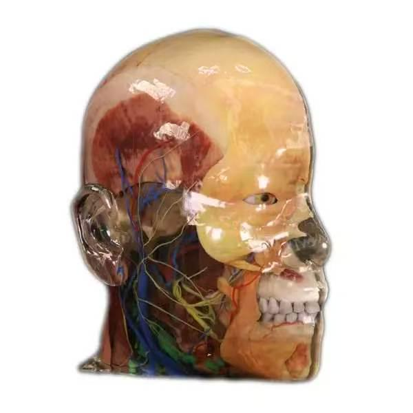
Recent advancements have allowed for the creation of multi-material replicas that reproduce anatomical geometry and selected material characteristics of human tissue with a high level of realism. This is achieved by extracting volume data from high-precision digital human data sets. For example, systems utilizing datasets with a voxel size of 0.0384mm by 0.0384mm by 0.1mm ensure that the resulting geometric models possess a visual and tactile perception consistent with formalin-fixed cadaveric specimens. This level of detail allows for a deeper exploration of intricate systems, such as vascular networks or nervous pathways, without the logistical and financial constraints associated with traditional cadaver labs.
Enhancing Surgical Planning and Medical Training
The application of these high-fidelity replicas extends far beyond the undergraduate anatomy classroom. In clinical environments, they serve as vital tools for both novice training and complex surgical preparation.
When preparing for complex surgical procedures, surgeons require more than just CT or MRI scans. They may use physical anatomical replicas to better understand complex anatomical structures and support simulation-based surgical training. This physical rehearsal can support procedural preparation and help surgical teams better understand complex anatomical relationships.
Furthermore, these tools facilitate improved communication across medical teams. By examining a precise physical replica, surgeons, radiologists, and nurses can collaborate more effectively during procedural planning. For institutions looking to integrate these advanced learning tools, exploring 3d printed anatomical models from DIGIHUMAN provides access to resources that combine multi-channel digital full-color printing with environmentally friendly resin materials, ensuring a 1:1 high simulation of physical anatomical structures.
Why Are 3D Printed Anatomical Models Becoming More Common in Medical Education?
The transition toward digital and physical anatomical replicas stems from several critical challenges in modern health sciences education. First, cadaver availability varies significantly among academic institutions worldwide due to strict regulatory frameworks, high ethical standards, and substantial maintenance costs. High-precision physical replicas offer a reliable, easily accessible alternative that ensures equal learning opportunities across all medical departments.
Additionally, these specialized models allow for repeated hands-on practice without physical degradation. Students can touch, inspect, and examine complex anatomical structures as many times as necessary to strengthen anatomical understanding. From an administrative perspective, these models function as standardized teaching materials. Instructors can deliver identical curriculum content to multiple training cohorts simultaneously, eliminating variables in tissue quality or dissection degradation.
Furthermore, these models foster interdisciplinary teaching. They enable medical, nursing, and biomedical engineering students to analyze identical spatial structures together during collaborative workshops. Finally, their durable construction guarantees long-term educational use, making them a cost-effective, multi-year investment for medical faculties worldwide.
Advantages Over Traditional Modalities
The integration of DIGIHUMAN‘s full-color, multi-material inkjet and light-curing printing technologies offers distinct advantages over older methods.
First, the customization offers greater flexibility for different teaching scenarios. Educators can specify models tailored to exact learning objectives—such as a heart model sectioned to display specific chamber pathologies, or a skull with detachable components to illustrate spatial relationships within the cranial cavity.
Second, the structural integrity and realism are vastly improved. Modern printers utilizing 12 or more material channels can combine single-hardness materials, soft and hard combinations, and transparent packaging within a single print. This means a single replica can accurately represent the rigidity of bone alongside the soft tissue of surrounding muscles and the delicate structure of intertwined blood vessels and nerves.
Finally, these replicas democratize access to high-quality anatomical resources. They provide a reproducible, cost-effective alternative that maintains a consistent standard of anatomical accuracy across different training cohorts, ensuring that all students and professionals have access to the tactile experience necessary for mastering human anatomy.
The Future of Interactive Medical Pedagogy
As digital modeling and manufacturing technologies continue to advance, their role in medical education will only expand. The ability to rapidly produce identical, high-precision copies of complex anatomical structures allows institutions to scale their hands-on training capabilities efficiently.
By moving beyond static, commercial models and embracing data-driven, multi-material replicas, medical schools and research hospitals are fostering a new standard of interactive learning. This commitment to advanced visualization modalities ensures that the next generation of healthcare professionals is equipped with a profound, practical understanding of the human body, ultimately leading to safer, more precise patient care.